Predictive Rule Extraction for Ocular Involvement in Behcet’s Disease
A Data Mining Approach Using Hospital-Based Data
DOI:
https://doi.org/10.22034/AIMI.2026.30.13Keywords:
Behçet’s Disease, Ocular Involvement , Data Mining , Decision Tree , Clinical Decision Support SystemsAbstract
Introduction: Ocular involvement is among the most severe and vision-threatening complications of Behçet’s disease (BD), making early risk stratification vital. This study aims to develop and validate an interpretable predictive data mining model to identify patients at high risk of ocular manifestations using a comprehensive hospital-based cohort.
Material and Methods: A retrospective analysis was conducted on clinical and demographic data from 1,613 patients diagnosed with Behçet’s disease at the rheumatology research center, Shariati hospital, Tehran. The J48 decision tree algorithm was implemented to extract explicit predictive rules. To establish a rigorous benchmark, a traditional logistic regression (LR) model was also evaluated. Both models were rigorously assessed using a 10-fold cross-validation framework across core metrics including accuracy, sensitivity, specificity, F1-score, and ROC-AUC.
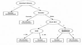
Results: The J48 decision tree model significantly outperformed the traditional logistic regression baseline, achieving a mean overall accuracy of 91.73±1.25, sensitivity of 74.78±1.84, specificity of 73.01±2.15 and AUC of 0.86±0.02 compared to an accuracy of 78.45 and an AUC of 74 for logistic regression. The root and primary nodes of the decision tree identified key risk factors, specifically genital ulceration, HLA-B51 status, sex, and ESR levels, generating transparent clinical rules for early identification.
Conclusion: The J48 decision tree algorithm provides superior predictive accuracy and clinical interpretability over conventional logistic regression for assessing ocular involvement in Behçet’s disease. These transparent rules offer a robust foundation for integration into Clinical Decision Support Systems (CDSS) to assist rheumatologists in proactive, individualized patient management.
References
1. Lavalle S, Caruso S, Foti R, Gagliano C, Cocuzza S, La Via L, et al. Behçet’s disease, pathogenesis, clinical features, and treatment approaches: A comprehensive review. Medicina (Kaunas). 2024; 60(4): 562. PMID: 38674208 DOI: 10.3390/medicina60040562
2. Fazaa A, Makhlouf Y, Ben Massoud F, Miladi S, Boussaa H, Ouenniche K, et al. Behçet disease: Epidemiology, classification criteria and treatment modalities. Expert Rev Clin Immunol. 2024; 20(12): 1437-48. PMID: 39101633 DOI: 10.1080/1744666X.2024.2388693
3. Aljohani R, Alharbi S. Epidemiology and clinical features of Behçet’s disease in the Middle East and North Africa: A systematic review and meta-analysis. Clin Rheumatol. 2026; 45(4): 2149-59. PMID: 41706278 DOI: 10.1007/s10067-026-07982-7
4. Kutlug Agackiran S, Kardes E, Agackiran A, Direskeneli H, Alibaz-Öner F. Common femoral vein wall thickness as a supportive imaging marker in the differential diagnosis of Behçet’s uveitis. Turk J Med Sci. 2026; 56(3): 761-8. PMID: 42499425 DOI: 10.55730/1300-0144.6209
5. Bozkaya Yücel B, Doğantan Ş, Çelik ÖG, Elmas S, Öğreten Yadigaroğlu N, Eser Öztürk H, et al. Clinical phenotype and long-term damage in paediatric Behçet’s disease: Comparative performance of ISG, ICBD and PEDBD classification criteria in a retrospective endemic cohort. RMD Open. 2026; 12(1): e006514. PMID: 41802833 DOI: 10.1136/rmdopen-2025-006514
6. Abdullah HA, Ahmed SA. Frequency of HLA-B* 51 in patients with Behcet’s disease and its relationship with the clinical findings in Kurdish population in Iraq. Genetics and Molecular Research. 2026; 25(8s): 1-8.
7. Vitale A, Caggiano V, Sbalchiero J, Lopalco G, Ragab G, Guerriero S, et al. The risk of developing ocular involvement among Behçet’s disease patients presenting with mucocutaneous involvement at disease onset: Data from the international AIDA network Behçet’s disease registry. Ophthalmol Ther. 2026; 15(1): 223-37. PMID: 41205142 DOI: 10.1007/s40123-025-01258-0
8. Nokhatha SAA, Aljaberi N, Zeyoudi JA, Almarzooqi A, Soukieh F, Khan M. High prevalence of HLA-B51 and ocular involvement in Behçet’s disease: A multicenter cross-sectional study. Rheumatol Int. 2025; 45(6): 140. PMID: 40349258 DOI: 10.1007/s00296-025-05892-6
9. Pei M, Qian Y, Liu X, Zhao C, Gao F, Zhang M. Visual prognosis of Behçet’s uveitis patients in China and its associated factors. Front Immunol. 2026; 17: 1617653. PMID: 41924254 DOI: 10.3389/fimmu.2026.1617653
10. Demiroğlu H, Barişta İ, Dündar S. Risk factor assessment and prognosis of eye involvement in Behçet's disease in Turkey. Ophthalmology. 1997; 104(4): 701-5. PMID: 9111266 DOI: 10.1016/s0161-6420(97)30249-8
11. Alalwany H, Alganmi N, Bawazir Y, Mustafa M, Abusamra H, Banjar H, et al. Enhancing the diagnosis of Behçet's disease using machine learning: A comparative study on clinical data from Saudi Arabia. Int J Telemed Appl. 2026; 2026: 6157852. PMID: 41583997 DOI: 10.1155/ijta/6157852
12. Qi S, Jiang Y, Li H, Gong X, Li Q, Zhou X, et al. Detection of rheumatoid arthritis-associated interstitial lung disease: A systematic review and meta-analysis. BMC Pulm Med. 2026; 26(1): 336. PMID: 42324528 DOI: 10.1186/s12890-026-04417-6
13. Fu D, Tan L, Yang Q. Machine learning in systemic lupus erythematosus research: A bibliometric and visual analysis (2015–2025). Egyptian Rheumatology and Rehabilitation. 2026; 53(1): 53.
14. Hammam N, Bakhiet A, El-Latif EA, El G, II, Samy N, Noor RAA, et al. Development of machine learning models for detection of vision threatening Behçet's disease (BD) using Egyptian college of rheumatology (ECR)-BD cohort. BMC Med Inform Decis Mak. 2023; 23(1): 37. PMID: 36803463 DOI: 10.1186/s12911-023-02130-6
15. Suwa A, Horita N, Ishido T, Takeuchi M, Kawagoe T, Shibuya E, et al. The ocular involvement did not accompany with the genital ulcer or the gastrointestinal symptoms at the early stage of Behçet’s disease. Mod Rheumatol. 2019; 29(2): 357-62. PMID: 29583036 DOI: 10.1080/14397595.2018.1457424
16. Seyahi E. Phenotypes in Behçet’s syndrome. Intern Emerg Med. 2019; 14(5): 677-89. PMID: 30747365 DOI: 10.1007/s11739-019-02046-y
17. Sada K-E, Miyawaki Y, Yanai R, Kida T, Onishi A, Yoshimi R, et al. Development and validation of data-driven, decision tree–based algorithms for identifying Behçet’s disease in claims data. Int J Med Inform. 2026; 209: 106266. PMID: 41506174 DOI: 10.1016/j.ijmedinf.2026.106266
18. Tangaro S, Lopalco G, Sabella D, Venerito V, Novielli P, Romano D, et al. Unraveling the microbiome-metabolome nexus: a comprehensive study protocol for personalized management of behçet’s disease using explainable artificial intelligence. Front Microbiol. 2024; 15: 1341152. PMID: 38410386 DOI: 10.3389/fmicb.2024.1341152
19. Hatemi G, Seyahi E, Fresko I, Talarico R, Uçar D, Hamuryudan V. Behçet’s syndrome: One year in review 2025. Clin Exp Rheumatol. 2025; 43(10): 1699-708. PMID: 41133359 DOI: 10.55563/clinexprheumatol/8xuim0
20. Sulu B, Karakoc A, Ak T, Dincer ZT, Senturk EF, Batur S, et al. Behçet’s syndrome within the spectrum of systemic vasculitides. Saudi J Ophthalmol. 2025; 39(4): 311-22. PMID: 41367840 DOI: 10.4103/sjopt.sjopt_355_24
21. Parsaei A, Moradi S, Masoumi M, Davatchi F, Najafi A, Kooshki AM, et al. Predictive value of erythrocyte sedimentation rate and C-reactive protein in Behcet's disease activity and manifestations: A cross-sectional study. BMC Rheumatol. 2022; 6(1): 9. PMID: 35144674 DOI: 10.1186/s41927-021-00241-z
Published
Issue
Section
License
Copyright (c) 2026 Niloufar Zaferani

This work is licensed under a Creative Commons Attribution 4.0 International License.
How to Cite