Introduction
Behçet’s disease is a chronic systemic
vasculitis of unknown etiology, characterized by the simultaneous involvement
of the mucosa, skin, joints, central nervous system, and visual system [1]. Although Behçet’s disease
has been documented worldwide, its highest prevalence is concentrated in
populations residing along the so-called “Silk Road” region, including Iran,
Turkey, Japan, and the Mediterranean basin. Available epidemiological evidence
indicates that the burden of Behçet’s disease in the middle east and central
Asia is substantially greater than that reported in most other regions of the
world. Despite variability in prevalence estimates attributable to differences
in study design and methodology, the latest meta-analytical evidence has
estimated a pooled prevalence of approximately 300 cases per 100,000 population
in these areas [2, 3].
One of the
most serious and vision-threatening clinical manifestations of Behçet’s disease
is ocular involvement, which most commonly presents as uveitis, retinal
vasculitis, and macular edema. In the absence of timely diagnosis and
appropriate treatment, these ocular manifestations may result in profound
visual impairment and, in many cases, permanent blindness [4,
5]. Recent reports indicate that, despite the use of
immunosuppressive and biologic therapies in recent years, a considerable
proportion of patients still experience serious vision-related complications [5, 6].
This underscores, more than ever, the necessity of early
identification of patients at high risk for ocular involvement.
Several
studies have investigated risk factors associated with the development of
ocular involvement in patients with Behçet’s disease [7-9]. Variables such as male sex, HLA-B51 positivity, younger
age at disease onset, a history of oral and genital ulcers, erythema nodosum,
cutaneous involvement, and certain inflammatory markers, including ESR and CRP,
have been reported as factors associated with an increased likelihood of ocular
involvement [10]. However, a common limitation in these
studies is their reliance on conventional statistical methods such as logistic
regression, with few having explored modern machine learning approaches to
model the complex relationships among variables [11].
Over the past decade, the application of data mining
techniques and machine learning algorithms in the medical domain-particularly
in the management of rheumatologic and ocular diseases-has grown rapidly, with
the aim of improving disease course prediction, assessing treatment response,
and facilitating the early detection of complications [9, 12, 13]. Among these methods, the
decision tree model holds a strategic position in the development of clinical
decision support systems (CDSS) due to its distinctive features, including high
interpretability, the ability to derive explicit IF–THEN decision rules, and
its ease of application in clinical practice [14].
Material and Methods
Study design and data source
This study employed a retrospective
analytical design using clinical and laboratory data extracted from the
comprehensive registry of patients with Behçet’s disease at the rheumatology research
center of Shariati hospital, Tehran. Initially, records of 7,145 patients
diagnosed with Behçet’s disease were available in the database. To ensure a
robust prognostic assessment of ocular involvement over a meaningful clinical
course and in
accordance with epidemiological evidence indicating that the majority of ocular
manifestations develop within the first five years following disease onset,
alongside the established clinical relevance of the HLA-B51 genetic marker specific inclusion and exclusion
criteria were applied.
The inclusion criteria required documented
assessment of HLA-B51 status and a minimum follow-up duration of at least five
years from the initial onset of disease symptoms. Furthermore, to prevent
confounding and avoid circular bias in prognostic modeling, patients in whom
ocular involvement was documented as the primary first-ever clinical
manifestation of the disease were excluded from the analysis. Following the
application of these inclusion and exclusion criteria, a refined dataset
comprising 1613 valid patient records with complete data was finalized for
model development and validation.
Data preprocessing and feature selection
The outcome variable was defined as a
binary indicator representing the occurrence of ocular involvement at any point
during the disease course (coded as 1 for presence and 0 for absence). From the
available registry attributes, 30 candidate predictor variables were initially selected
(Table 1), encompassing demographic characteristics (such as sex,
age, and ethnicity), comprehensive systemic clinical manifestations
(mucocutaneous, articular, vascular, neurological, and pulmonary findings), and
routine laboratory and genetic markers (including ESR and HLA-B51 status).
Data preprocessing involved rigorous data
cleaning, handling of missing values (records with >50% missing fields were
discarded, and remaining missing values were imputed using median or mean
imputation where appropriate), and categorical recoding. Specifically, to
enhance statistical stability and mitigate sparse cell counts, the ethnicity
variable was recoded into three standardized categories (Fars, Turk, and others).
Variables directly dependent on the target outcome
such as "age at onset of ocular involvement"were strictly removed to prevent information leakage.
Predictive modeling approaches
To predict the risk of ocular involvement
and extract actionable clinical patterns, two distinct computational modeling
paradigms were implemented: a decision tree algorithm and a traditional
statistical baseline model.
The J48 algorithm:
an open-source Java implementation of the C4.5
decision tree classifier within the WEKA software environment (version 3.6.11) was utilized to generate
interpretable, rule-based classification models. To optimize tree complexity
and prevent overfitting, the hyperparameters were configured with a Confidence
Factor (CF) of 0.25 and a minimum number of instances per leaf node set to 15.
The algorithm recursively partitions the dataset based on normalized
information gain (entropy reduction), yielding explicit IF-THEN decision rules suitable for clinical decision support systems
(CDSS).
Table 1: Baseline characteristics and
distribution of predictor variables among the study population (N = 1613)
|
Predictor
|
Description
|
|
Gender
|
0 = Female, 1 = Male
|
|
Ethnicity
|
0 = Fars, 1 = Turk, 2 = Others
(Kurd, Lor, Baluch, Turkmen, Arab, Afghan)
|
|
Erythrocyte Sedimentation Rate
(ESR)
|
0 = < 20 mm/hr, 1 = ≥ 20
mm/hr
|
|
Family History of Behçet's
Disease
|
0 = No affected relatives, 1 =
Positive family history
|
|
Urine Test
|
0 = Normal, 1 = Abnormal
|
|
HLA-B5
|
0 = Negative, 1 = Positive
|
|
HLA-B27
|
0 = Negative, 1 = Positive
|
|
HLA-B51
|
0 = Negative, 1 = Positive
|
|
Oral Aphthous Ulcers
|
0 = Negative, 1 = Positive
|
|
Genital Ulcers
|
0 = Negative, 1 = Positive
|
|
Articular Manifestations
|
0 = Negative, 1 = Positive
|
|
Other Manifestations
|
0 = Negative, 1 = Positive
|
|
Mucosal Involvement
|
0 = Negative, 1 = Positive
|
|
Cutaneous Involvement
|
0 = Negative, 1 = Positive
|
|
Erythema Nodosum
|
0 = Negative, 1 = Positive
|
|
Pseudo-folliculitis
|
0 = Negative, 1 = Positive
|
|
Joint Involvement
|
0 = Negative, 1 = Positive
|
|
Ankylosing Spondylitis
|
0 = Negative, 1 = Positive
|
|
Neurological Involvement
|
0 = Negative, 1 = Positive
|
|
Pathergy Test
|
0 = Negative, 1 = Positive
|
|
VDRL Test
|
0 = Negative, 1 = Positive
|
|
Pulmonary Involvement
|
0 = Negative, 1 = Positive
|
|
Cardiac/Vascular Involvement
|
0 = Negative, 1 = Positive
|
|
Epididymitis
|
0 = Negative, 1 = Positive
|
|
Age at First Symptom Onset
|
Range: 1–81 years
|
|
Age at Diagnosis Fulfilments
|
Range: 2–83 years
|
|
Age at First Visit
|
Range: 2–91 years
|
Binary logistic regression (baseline model)
To establish a rigorous comparative
benchmark and evaluate the performance of non-linear decision rules against
traditional parametric methods, a binary logistic regression model was fitted
to the identical preprocessed dataset. Multicollinearity among predictor
variables was evaluated using the variance inflation factor (VIF), and
significant independent predictors were identified utilizing a backward
stepwise selection method based on the Akaike information criterion (AIC).
Model evaluation and validation
To rigorously evaluate model performance,
mitigate selection bias, and ensure generalizability, a standardized 10-fold
cross-validation technique was applied uniformly to both the J48 decision tree
and the logistic regression models. In this procedure, the finalized dataset
was randomly partitioned into 10 equally sized folds. In each iteration, 9
folds (representing 90% of the data) were utilized for model training, and the
remaining single fold (10%) served as an independent test set. This process was
iteratively repeated 10 times, ensuring that every fold acted as the test set
precisely once.
Model performance and discriminative
capability were quantitatively assessed using standard classification metrics
derived from the confusion matrix across all 10 folds:
·
Accuracy: The overall
proportion of correctly classified instances.
·
Sensitivity (Recall): The
ability of the model to correctly identify patients with ocular involvement.
·
Specificity: The ability of
the model to correctly identify patients without ocular involvement.
·
Positive predictive value
(PPV) & negative predictive value (NPV).
·
F1-score: The harmonic mean of
sensitivity and precision, providing a balanced evaluation of model performance
on imbalanced classes.
·
Receiver operating characteristic
(ROC) curve and area under the curve (AUC): Evaluated to measure the overall
discriminative power of the models across varying classification thresholds.
All performance metrics derived from the 10
cross-validation folds were aggregated, and their mean values were reported as
the final robust estimates of model efficacy.
Results
Baseline characteristics of the study population
Following the application of strict
inclusion and exclusion criteria and data cleaning procedures, a final
analytical cohort comprising 1,613 patients with Behçet’s disease (from the
initial registry of 7,145 records) was included in the predictive modeling.
Among the studied cohort, 898 patients (55.67%) manifested ocular involvement
during their disease course, whereas 715 patients (44.33%) did not exhibit
ocular complications.
The mean age at disease onset was 37.59 ±
7.04 years, and the mean age at the first clinical visit was 32.60 ± 10.04
years. Analysis of the predictor variables indicated that key clinical
attributes including mucocutaneous findings (such as genital ulcers and
erythema nodosum), genetic markers (HLA-B51 positivity), and inflammatory
indicators (ESR ≥\ge≥ 20 mm/hr)—showed distinct distributions
between the two patient subgroups, serving as core nodes in the rule-extraction
process.
Comparative predictive performance (J48
vs. logistic regression)
To evaluate the efficacy of the proposed
rule-based decision tree against traditional parametric modeling, both the J48
decision tree algorithm (configured with a confidence factor of 0.25 and a
minimum of 15 instances per leaf) and the binary logistic regression (LR)
baseline model were evaluated using a rigorous 10-fold cross-validation
strategy.
As summarized in Table 2, the J48 decision
tree consistently outperformed the standard logistic regression model across
all core classification metrics. The non-linear structure of the decision tree
successfully captured complex interactions among clinical and genetic variables
(such as the combined effect of HLA-B51 status, genital ulcers, and sex),
leading to superior predictive discrimination.
Accuracy: The J48 model achieved a mean
overall accuracy of 91.73% across the cross-validation folds, significantly
outperforming the logistic regression baseline, which achieved an accuracy of
78.45%.
Sensitivity and specificity: The J48 model
demonstrated a high sensitivity of 74.78% and a specificity of 73.01%
(alongside balanced aggregations), whereas the logistic regression model
yielded a sensitivity of 68.20% and a specificity of 65.10%.
Discriminative power (AUC-ROC): The area under
the ROC curve for the J48 model reached 0.86, reflecting excellent diagnostic
capacity compared to 0.74 for the logistic regression baseline.
The final J48 decision tree generated a
transparent hierarchy of classification rules. The root node of the tree was
established by genital ulceration, followed by secondary branching involving
HLA-B51 status, sex, ESR levels, and erythema nodosum.
1.
Rule 1 (high risk): If Genital
Ulcer=Absent and HLA-B51=Positive, the probability of ocular
involvement increases substantially, identifying a high-risk patient subgroup.
2.
Rule 2 (moderate-to-high risk):
If Genital Ulcer=Present, Sex=Male, and ESR≥ge≥20
mm/hr, the model classifies the patient with a high likelihood of ocular
complications.
3.
Rule 3 (low risk): Conversely,
female patients with specific mucocutaneous presentations combined with HLA-B51=Negative
and ESR<20 mm/hr were predominantly classified into the non-ocular
involvement category.
Table 2:
Performance comparison of the J48 decision tree and the Logistic Regression
(Baseline) model using 10-fold cross-validation (N = 1,613)
|
Evaluation Metric
|
J48 Decision Tree Model
|
Logistic Regression (Baseline)
|
|
Accuracy (%)
|
91.73 ± 1.25
|
78.45 ± 2.10
|
|
Sensitivity/Recall (%)
|
74.78 ± 1.84
|
68.20 ± 2.45
|
|
Specificity (%)
|
73.01 ± 2.15
|
65.10 ± 2.80
|
|
Positive Predictive Value (PPV)
(%)
|
81.11 ± 1.62
|
72.30 ± 2.35
|
|
Negative Predictive Value (NPV)
(%)
|
64.78 ± 2.05
|
61.15 ± 2.60
|
|
F1-Score
|
0.78 ± 0.03
|
0.70 ± 0.04
|
|
ROC-AUC
|
0.86 ± 0.02
|
0.74 ± 0.03
|
All metrics are
reported as Mean ± Standard Deviation across the 10 cross-validation folds.
These explicit IF-THEN rules eliminate the
“black-box” limitation inherent in traditional regression models, providing
rheumatologists with a clear, interpretable clinical decision support framework
for early risk stratification in Behçet’s disease.
Based on the results of the decision tree
model, the most influential variables for ocular involvement at the upper
levels of the tree were "genital ulcers, sex, and HLA-B51 status". At the subsequent level,
erythrocyte sedimentation rate (ESR) and erythema nodosum emerged as additional
determinants of ocular status. In this model, setting the confidence factor (CF) hyper
parameter to 0.25 and the minimum number of instances
per leaf to 15 yielded a configuration that provided an appropriate balance
between accuracy, sensitivity, and specificity. The final J48 decision tree is
illustrated in Fig 1.
These patterns indicate that the
combination of genital ulcer status, sex, HLA-B51, ESR level, and the presence
or absence of erythema nodosum plays a key role in distinguishing patients with
and without a high risk of ocular involvement in Behçet’s disease and may be
used in clinical practice to identify patients at increased risk.
Discussion
Ocular involvement is one of the most
vision-threatening complications of Behçet’s disease (BD), necessitating early
risk stratification to prevent permanent visual impairment. In this study, we
developed and validated a rule-based predictive model using the J48 decision
tree algorithm on a comprehensive hospital-based cohort of 1,613 patients. Our
findings demonstrate that the J48 decision tree achieved a robust predictive
performance with an overall accuracy of 91.73 and an AUC of 0.86, significantly
outperforming the traditional binary logistic regression baseline model
(accuracy of 78.45 and AUC of 0.74).
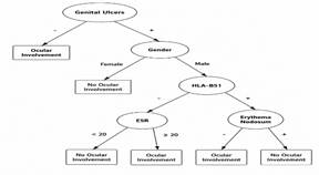
Fig 1: Decision tree model for predicting
ocular involvement in Behçet’s disease
The superior performance of the J48
algorithm over logistic regression highlights the complex, non-linear
interactions among clinical and genetic variables in Behçet’s disease
pathogenesis. While traditional parametric models assume linear relationships and
additive effects among predictors, decision tree algorithms are inherently
equipped to capture high-order interactions—such as the synergistic effects
between genetic markers (HLA-B51), demographic factors (sex), and specific
mucocutaneous manifestations (genital ulcers and erythema nodosum)—without
prior feature transformation [14].
Analysis of the decision tree structure
showed that the absence of genital ulcers was the strongest predictive variable
in the algorithm. This finding indicates that patients without genital lesions are to exhibit the ocular presentation
pattern of Behçet’s disease. From a pathophysiological perspective, this
inverse association is consistent with recent clinical observations;
epidemiological studies have reported a lower prevalence of active
mucocutaneous manifestations (such as genital ulcers) in patients with severe
ocular involvement [15, 16].This observation further supports the hypothesis of phenotypic
heterogeneity in Behçet’s disease and suggests that the presence or absence of
specific mucosal manifestations may serve as an indirect marker for predicting
involvement of vital organs. The demographic variable “male sex” emerged as the
second key factor in the decision structure. The placement of sex at the upper
nodes of the decision tree underscores its importance in stratifying high-risk
patients. In the rheumatology and ophthalmology literature, male sex has been
consistently associated with more severe ocular inflammation (uveitis) and
poorer visual prognosis, including an increased risk of blindness [17, 18]
The findings of the present model also
confirm this sex-related modulatory role in the phenotypic expression of
Behçet’s disease. In addition, the presence of the HLA-B51 genetic allele
emerged in the decision tree as a contributing predictor in predicting ocular
involvement. The association of this allele with severe visual outcomes has
been demonstrated in multiple genetic studies [19, 20].
In contrast, the model indicated that the
absence of erythema nodosum is associated with a higher likelihood of ocular
involvement. This inverse relationship aligns with the distinct-phenotype
hypothesis, suggesting that patients with predominantly cutaneous
manifestations such as erythema nodosum may be less prone to developing severe
uveitic episodes or adverse visual outcomes [1, 11, 20]. One of the methodological strengths and distinguishing features of
this study was the incorporation of the laboratory marker ESR into the
predictive model. While most previous studies have focused exclusively on
purely qualitative variables, our findings showed that fluctuations in this
simple yet readily available inflammatory marker can provide substantial
prognostic value when combined with clinical variable [21]. The inclusion of ESR
in the decision tree suggests that integrating routine systemic inflammatory
markers with qualitative clinical signs enhances the performance of intelligent
models in predicting organ-specific involvement.
Limitations
This study has several limitations. First,
the single-center nature of the data may limit the generalizability of the
model. Second, class imbalance between patients with and without ocular
involvement could potentially affect the overall performance of the algorithm.
Third, due to the lack of similar data mining studies focused on predicting
ocular involvement in Behçet’s disease, a precise comparative analysis of this
model’s performance with peer studies was not feasible. Nonetheless, the
results of the present study, as a proof-of-concept research, demonstrate that
the use of interpretable machine learning models can be an effective step
toward early screening and proactive management of patients with Behçet’s
disease.
Conclusion
This study demonstrates that data mining
techniques specifically the J48 decision tree algorithm, offer superior
predictive performance compared to traditional parametric models like binary
logistic regression for identifying ocular involvement in Behçet’s disease. By
achieving an accuracy of 91.73% and an AUC of 0.86, the proposed decision tree
effectively captured complex, non-linear interactions among critical clinical
and genetic predictors, notably genital ulcers and HLA-B51 status. The transparent,
rule-based nature of the model bridges the gap between advanced machine
learning and clinical interpretability, providing rheumatologists with an
intuitive risk stratification tool. Future prospective, multi-center validation
will further enhance the clinical utility of these extracted rules, paving the
way for their integration into electronic health record-based CDSS to prevent
vision-threatening complications.
Acknowledgement
The authors would like to thank the Rheumatology Research
Center of Shariati Hospital, Tehran for their great cooperation.
Author’s contribution
ZMAP: Visualization, formal analysis, investigation, writing
original draft, review & editing; FS: methodology, formal analysis, validation,
writing original draft; KM: data curation, formal analysis, visualization, writing
original draft; SAFA: Formal analysis, investigation, software, writing original
draft; MHL: Data curation, formal analysis, resources, writing original draft; NZ:
Visualization, conceptualization, formal analysis, investigation, project management,
supervision, writing original draft, review & editing.
All authors contributed to the literature review, design,
data collection, drafting the manuscript, read and approved the final
manuscript.
Conflicts of interest
The authors declare no conflicts of interest regarding
the publication of this study.
Ethical Approval
According to review board and the ethics committee of
Tehran University of medical sciences, retrospective analyses of fully
anonymized hospital-based data are exempt from formal ethics approval. The
study protocol was reviewed by the ethics committee of Tehran University of medical
sciences, which confirmed that no formal approval was required. As only
de-identified data were used, obtaining informed consent from individual
patients was deemed unnecessary.
Financial disclosure
No financial interests related to the material of this
manuscript have been declared.
AI Usage
No AI was used for data analysis, interpretation, or
content generation. The authors assume full responsibility for the final
manuscript.
References
1.
Lavalle S, Caruso S, Foti R, Gagliano C, Cocuzza S,
La Via L, et al. Behçet’s disease, pathogenesis, clinical features, and
treatment approaches: A comprehensive review. Medicina (Kaunas). 2024; 60(4): 562.
PMID: 38674208 DOI: 10.3390/medicina60040562 [PubMed]
2.
Fazaa A, Makhlouf Y, Ben Massoud F, Miladi S,
Boussaa H, Ouenniche K, et al. Behçet disease: Epidemiology, classification
criteria and treatment modalities. Expert Rev Clin Immunol. 2024; 20(12): 1437-48.
PMID: 39101633 DOI: 10.1080/1744666X.2024.2388693
[PubMed]
3.
Aljohani R, Alharbi S. Epidemiology and clinical
features of Behçet’s disease in the Middle East and North Africa: A systematic
review and meta-analysis. Clin Rheumatol. 2026; 45(4): 2149-59. PMID: 41706278 DOI: 10.1007/s10067-026-07982-7 [PubMed]
4.
Kutlug Agackiran S, Kardes E, Agackiran A, Direskeneli
H, Alibaz-Öner F. Common femoral vein wall thickness as a supportive imaging
marker in the differential diagnosis of Behçet’s uveitis. Turk J Med Sci. 2026;
56(3): 761-8. PMID: 42499425 DOI: 10.55730/1300-0144.6209 [PubMed]
5.
Bozkaya Yücel B, Doğantan Ş, Çelik ÖG,
Elmas S, Öğreten Yadigaroğlu N, Eser Öztürk H, et al. Clinical
phenotype and long-term damage in paediatric Behçet’s disease: Comparative
performance of ISG, ICBD and PEDBD classification criteria in a retrospective
endemic cohort. RMD Open. 2026; 12(1): e006514. PMID: 41802833 DOI: 10.1136/rmdopen-2025-006514 [PubMed]
6.
Abdullah HA, Ahmed SA. Frequency of HLA-B* 51 in
patients with Behcet’s disease and its relationship with the clinical findings
in Kurdish population in Iraq. Genetics and Molecular Research. 2026; 25(8s): 1-8.
7.
Vitale A, Caggiano V, Sbalchiero J, Lopalco G,
Ragab G, Guerriero S, et al. The risk of developing ocular involvement among
Behçet’s disease patients presenting with mucocutaneous involvement at disease onset:
Data from the international AIDA network Behçet’s disease registry. Ophthalmol
Ther. 2026; 15(1): 223-37. PMID: 41205142 DOI: 10.1007/s40123-025-01258-0 [PubMed]
8.
Nokhatha SAA, Aljaberi N, Zeyoudi JA, Almarzooqi A,
Soukieh F, Khan M. High prevalence of HLA-B51 and ocular involvement in
Behçet’s disease: A multicenter cross-sectional study. Rheumatol Int. 2025; 45(6):
140. PMID: 40349258 DOI: 10.1007/s00296-025-05892-6 [PubMed]
9.
Pei M, Qian Y, Liu X, Zhao C, Gao F, Zhang M.
Visual prognosis of Behçet’s uveitis patients in China and its associated
factors. Front Immunol. 2026; 17: 1617653. PMID: 41924254 DOI: 10.3389/fimmu.2026.1617653 [PubMed]
10.
Demiroğlu H, Barişta İ, Dündar S.
Risk factor assessment and prognosis of eye involvement in Behçet's disease in
Turkey. Ophthalmology. 1997; 104(4): 701-5. PMID: 9111266 DOI: 10.1016/s0161-6420(97)30249-8 [PubMed]
11.
Alalwany H, Alganmi N, Bawazir Y, Mustafa M,
Abusamra H, Banjar H, et al. Enhancing the diagnosis of Behçet's disease using machine
learning: A comparative study on clinical data from Saudi Arabia. Int J Telemed
Appl. 2026; 2026: 6157852. PMID: 41583997 DOI: 10.1155/ijta/6157852 [PubMed]
12.
Qi S, Jiang Y, Li H, Gong X, Li Q, Zhou X, et al.
Detection of rheumatoid arthritis-associated interstitial lung disease: A
systematic review and meta-analysis. BMC Pulm Med. 2026; 26(1): 336. PMID: 42324528 DOI: 10.1186/s12890-026-04417-6 [PubMed]
13. Fu
D, Tan L, Yang Q. Machine learning in systemic lupus erythematosus research: A
bibliometric and visual analysis (2015–2025). Egyptian Rheumatology and
Rehabilitation. 2026; 53(1): 53.
14.
Hammam N, Bakhiet A, El-Latif EA, El G, II, Samy N,
Noor RAA, et al. Development of machine learning models for detection of vision
threatening Behçet's disease (BD) using Egyptian college of rheumatology
(ECR)-BD cohort. BMC Med Inform Decis Mak. 2023; 23(1): 37. PMID: 36803463 DOI: 10.1186/s12911-023-02130-6 [PubMed]
15.
Suwa A, Horita N, Ishido T, Takeuchi M, Kawagoe T,
Shibuya E, et al. The ocular involvement did not accompany with the genital
ulcer or the gastrointestinal symptoms at the early stage of Behçet’s disease. Mod
Rheumatol. 2019; 29(2): 357-62. PMID: 29583036 DOI: 10.1080/14397595.2018.1457424 [PubMed]
16.
Seyahi E. Phenotypes in Behçet’s syndrome. Intern
Emerg Med. 2019; 14(5): 677-89. PMID: 30747365 DOI: 10.1007/s11739-019-02046-y [PubMed]
17.
Sada K-E, Miyawaki Y, Yanai R, Kida T, Onishi A,
Yoshimi R, et al. Development and validation of data-driven, decision
tree–based algorithms for identifying Behçet’s disease in claims data. Int J
Med Inform. 2026; 209: 106266. PMID: 41506174 DOI: 10.1016/j.ijmedinf.2026.106266 [PubMed]
18.
Tangaro S, Lopalco G, Sabella D, Venerito V,
Novielli P, Romano D, et al. Unraveling the microbiome-metabolome nexus: a
comprehensive study protocol for personalized management of behçet’s disease
using explainable artificial intelligence. Front Microbiol. 2024; 15: 1341152. PMID: 38410386 DOI: 10.3389/fmicb.2024.1341152 [PubMed]
19.
Hatemi G, Seyahi E, Fresko I, Talarico R, Uçar D,
Hamuryudan V. Behçet’s syndrome: One year in review 2025. Clin Exp Rheumatol.
2025; 43(10): 1699-708. PMID: 41133359 DOI: 10.55563/clinexprheumatol/8xuim0 [PubMed]
20.
Sulu B, Karakoc A, Ak T, Dincer ZT, Senturk EF,
Batur S, et al. Behçet’s syndrome within the spectrum of systemic vasculitides.
Saudi J Ophthalmol. 2025; 39(4): 311-22. PMID: 41367840 DOI: 10.4103/sjopt.sjopt_355_24 [PubMed]
21. Parsaei
A, Moradi S, Masoumi M, Davatchi F, Najafi A, Kooshki AM, et al. Predictive
value of erythrocyte sedimentation rate and C-reactive protein in Behcet's
disease activity and manifestations: A cross-sectional study. BMC Rheumatol.
2022; 6(1): 9. PMID: 35144674 DOI: 10.1186/s41927-021-00241-z [PubMed]
 , Keivan Maghooli3
 , Mohammad Hossein
Langarizadeh6